Regency Medical Centre’s Radiology and Specialised Diagnostic Services wing is packed with the latest in imaging technology. The hospital specialises in a range of diagnostic radiology.
Radiology is the branch of medicine that deals with technology relating to the imaging of the human body. This imaging is utilized by doctors to pin-point the problem areas and causes for a variety of diseases.
The doctors at RMC are some of the most proficient and highly trained in the country. Moreover, they are further empowered by some of the latest in imaging technology at hand.
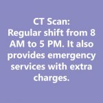
SPIRAL High Resolution CT Scan

This procedure uses a computer that is linked to an X-ray machine and it takes a series of pictures of specified areas of the human body.The machine scans the body on a spiral path, thus giving it its
name. This is a great means of providing the treating physician with a higher number of images in a considerably shorter duration of time.
Computed Tomography, or CT scan, uses x-rays to create a cross-sectional image of the body/body part. At times a contrast dye is used to enhance the
structures or fluids in the body.
This test can be used to diagnose an infection, guide a surgeon to the right area, identify tumours and masses, and to study blood vessels. Once the images are taken the computer creates separate images of the body, these are called slices.
These can be viewed on a computer, in print or even stored for later use. At RMC we perform all kinds of scans and angiograms in our CT scan department like coronary, pulmonary, peripheral angio, etc.
Doctors reporting on these scans are experts both local and international to provide our patients the highest standards of
service.
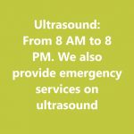
Ultrasound

Ultrasound has been one of the most popular imaging technologies used in medicine for years. This technology uses high frequency sound waves and their echoes to create an image of a specified organ.
This method is highly reminiscent of that used by bats and dolphins, known as echolocation.
The reflected sound waves create a picture for the doctor to analyse. These high frequency sound waves travel at 1 to 5 megahertz with the help of probe.
These sound waves hit a boundary between tissues and some of them are reflected back. These reflected waves are picked up by the probe and
relayed to the machine which creates the image.
The machine measures the distance of the organ and/or tissue from the probe. The image generated comes with all this information written out for
the doctor to peruse.
Ultrasound For visceral Organs

Ultrasound for the visceral organs ensures that an image is created of the structures within the body like, kidneys, liver, gall bladder, bile ducts, pancreas, spleen and abdominal aorta.
The transducer can be moved along the surface of the skin to create image of the body parts that need to be seen.
There is hardly any preparation required for getting an ultrasound. The doctor will inform you if you need to fast before the test. This test is used to diagnose a number of conditions like:
- Abdominal pain or distention
- Abnormal liver function
- Enlarged abdominal organ
- kidney stones, gallstones
- or even an abdominal aortic aneurysm.
Ambulatory BP

Ambulatory Blood Pressure Measurement or ABPM, is the process of measuring blood pressure at regular intervals. This alleviates the sense of nervousness most people feel in hospitals as the measurement is done on out-of- office.These kind of measurements are considered very important by most hypertension organisations. This kind of blood pressure measurement allows to monitor the patient for long durations.
The readings from ambulatory BP can also help measure the impact of
hypertension on various organs in the body. It is also a great way to check overnight surges in BP or nocturnal dips in the pressure.
RMC uses the best machines and equipment possible to make sure
that we are getting the most accurate readings possible for your ambulatory BP test.
TMT

The treadmill test, also known as the exercise stress test or the computerised stress test, is usually performed to check the severity of heart disease.
This test measures the effect of physical stress on your heart. These tests can detect flow-limiting blockages in the coronary arteries. This test is usually
done to check if an asymptomatic person has coronary artery disease.
It is also used to check the cause for symptoms like tightness of chest, difficulty in breathing etc.
In patients who are on medicines for blocked arteries this test can be used to check their progress. It is also crucial to check a person’s exercise tolerance before beginning exercise or cardiac rehabilitation program.
This test is performed with a continuous monitoring on the ECG while the person runs or walks on a treadmill. A person giving the TMT test must not eat or drink 6 hours prior to the test. If you are on medications
your doctor will advise you if you need to skip any.
Ambulatory Blood Pressure Measurement or ABPM, is the process of measuring blood pressure at regular intervals. This alleviates the sense of nervousness most people feel in hospitals as the measurement is done on out-of- office.These kind of measurements are considered very important by most hypertension organisations. This kind of blood pressure measurement allows to monitor the patient for long durations.
The readings from ambulatory BP can also help measure the impact of
hypertension on various organs in the body. It is also a great way to check overnight surges in BP or nocturnal dips in the pressure.
RMC uses the best machines and equipment possible to make sure
that we are getting the most accurate readings possible for your ambulatory BP test.
Audiometry

Audiometry tests are used to determine the patient’s hearing abilities. This test uses an audiometer to test the subject’s hearing levels and acuity of perceiving various sounds, intensity and pitch, among other things.
This test checks for a complete range of inner ear functions. The audiometer is a machine that plays sounds through the headphones and the audiologist will be guiding you through the test.
Various tests are administered in the audiometry test including the tuning fork test. There will be separate tests for each test that will be administered in order to determine the loss of hearing and the extent of it.
Colposcopy

This test is a means of examining the cervix, vagina and vulva with an medical instrument known as the colposcope.
This test is usually performed if an individual’s Pap smear test has shown unusual results. The colposcope is essentially an electric microscope with a light that enables the doctor to view the cervix in a magnified fashion to check for abnormalities.
This test lasts for about 15 to 20 minutes and does not require any anaesthesia.
The colposcope does not touch you and takes photographs of area that the doctor deems suspicious. To aid this a speculum is used to hold the walls of the vagina open for the doctor to see your cervix.
A biopsy may be conducted of areas that are deemed suspicious by the doctor.
Uroflowmetry

This test is used for checking the amount of urine voided at a time as well as the speed of urination.
It is an essential test for doctors to check the causes of certain urinary issues.
This test is usually recommended for people with issues like slow urination, weak urine stream, or have difficulty urinating.
It can also be used to test the sphincter muscle. This test tells the doctor how well your bladder and sphincter are functioning and can help the doctor estimate the blockage or obstruction,
if any.
This test can also help identify other problems like an enlarged prostate or even a weakened bladder. Make sure you arrive for the test with a full bladder.
This test requires you to urinate in a funnel shaped device or a special toilet. An electric uroflowmeter is attached to the toilet or funnel to derive the readings required by the doctor.
X-ray Machine

This is one of the most widely used imaging machine in medicine. X-rays are highly penetrative, ionizing radiation.
X-ray machines are used to create images of dense tissues like bones and even teeth. The x-rays pass through the body, and on to a photographic plate.
Since the x-rays pass through the soft tissue far more easily and are absorbed far more by the dense tissue like bones, it creates an image of the body part for the doctor to examine.
We, at RMC, provide all possible x-ray options including, barium meal, barium enema, barium follow through, barium swallow, IVU and urethrogram, among others.
Echocardiography & Colour Doppler

This procedure uses Doppler ultrasonography to examine the heart and its functioning. The echocardiogram makes use of the high frequency sound waves to create an image of the heart while using the Doppler technology to judge the speed and direction of the flow of the blood.This technology allows for a highly accurate assessment of the flow of the blood and cardiac tissue at any point.
This procedure is also widely used because of the ease it provides to measure the flow of blood within the heart without needing invasive procedures.
Contrast enhanced ultrasound can be used to improve velocity or other flow related medical measurements.
GE Echo Machine for Echocardiography

GE’s Echocardiography machine is one of the finest available. The machine works on the simple theory of imaging using sound. The machine uses high frequency sound waves to create a picture of the patient’s heart to pinpoint the trouble they may be facing.
This test is also known as echocardiography or diagnostic cardiac ultrasound.
This test is used in order to see the size and shape of the heart and identify the thickness and movements of your heart’s walls.
It is used to check on factors like the heart’s pumping strength, how your heart moves, if the heart valves are functioning correctly, if the blood is leaking backward through your heart valves or if your heart valves are too narrow. This test does not hurt at all and has absolutely no side effects.
Holter Monitor

A Holter monitor is a battery-operated portable device that is used to check the heart’s activity for the period of 24 to 48 hours. The size of the device is about that of a small camera and has wires and electrodes that are attached to your skin.
This device measures and records your ECG as you go about your daily activity. If one has a slow, fast or irregular heart beat they may be asked to wear a Holter monitor.
The regular electrocardiograms help the doctor understand and analyse your heart’s activity thoroughly. There are no risks to wearing this monitor and it causes no pain to the patient.
The patient may be required to keep a diary of their activities for the doctor to refer to while checking the report. Your diary’s entries will be used to compare with the changes in your heart patterns.
EGG

This is an electrophysiological monitoring method to record essential electrical activity of the brain.
A non-invasive process, EEG monitors the fluctuations that arise because of the ionic current within the neurons of the brain.
Clinically speaking, the EEG records the brain’s spontaneous electrical
activity over a period of time. The procedure involves placing a number of electrodes in contact with the scalp.
Each electrode is connected to a differential amplifier. EEG systems these days are digital and the screen displays the different brainwave readings.
This system is used to diagnose sleep disorders, depth of anaesthesia, coma, encephalopathies and brain death.
It is also used to diagnose epilepsy and its effect on the brain. Doctors reporting on the EEGs are specialists and experts, both
local and from around the world in order to give our patients the best possible treatment.
Tympanometry

The tympanic membrane inside the ear is crucial not only for our ability to hear but also for our balance.
It separates the middle ear from the outer ear. The results of the test are recorded on a graph called the tympanogram.
Through this test the doctor can check for fluid in the middle ear, middle ear infection, perforation or tear in the ear drum, or any problems with the Eustachian tube.
The doctors first check your ear with an otoscope to ensure the passage of the ear is clear. A probe type device is then placed at the entrance of the ear.
One may feel a little discomfort when the device changes the pressure in your ear to check the movement of the eardrum and takes reading.
One is not allowed to move, speak or swallow during the test as it may interfere with the readings.
Colonoscopy

This test is used by doctors to evaluate and check the inside of the colon. The instrument used for this procedure is called a colonoscope.
It is a four foot long, flexible tube, which is about the thickness of a finger. It has a camera and a light at the end of it which enables the doctors to get a
clear, magnified view.
The colonoscope is inserted from the anus and is advanced slowly with full
visual control through the colon and as far as the rectum.
Colonoscopies are usually done as a test for colon cancer, to identify cause for blood in stool, diarrhoea, abdominal pain, change in bowel habit, or abnormalities found in colonic x-rays and CT scans. This process requires the patient to have an absolutely clean bowel over and above a number of special preparations.
The doctors will give detailed instructions about achieving clean bowels and what is required off the patients.
Gastrointestinal Scopes

Gastrointestinal endoscopy is a procedure that allows the doctor to view the inner lining of the digestive tract.
RMC has a range of gastrointestinal scopes to allow the doctors a range of options to provide patients the best possible care. The endoscope is essentially a tube with a camera and light fibres at one end.
Sigmoidoscopy is a procedure where a doctor uses a specialised endoscope to check your large intestine. This is the best possible means to check for colon cancer and can be used to investigate a number of problems.
Pulmonary Function Tests (PFT)

This test is used to measure how well the patient’s lungs are functioning. The Pulmonary Function Test, or PFT, checks how well an individual is breathing and the efficacy of the lungs in providing oxygen to the rest of the body.
This test is ordered if the patient presents with symptoms of lung
problems, if they are exposed to dangerous substances at their workplace, to monitor chronic lung disease, or to assess lung capacity after surgery.
Tests may include spirometry where you are asked to breathe in and out in front of a machine fitted with a mouthpiece.
The technician will explain how you need to breathe during the test. A plethysmography test measures the volume of the gas in your lungs, it is also known as lung volume.
There is also a diffusion capacity test to test how the alveoli in the lungs work. One should avoid having a big meal before PFTs and foods containing caffeine should also be avoided.
Avoid smoking at least an hour before the test.


If you have diabetes this test needs to be done every 2 to 6 months. This test measure your average blood sugar level and because of this it is not necessary to fast for a day.
X-rays during pregnancy do not increase chances of miscarriage or cause problems to the unborn child.However, repeated exposure to radiation can damage the cells of the body leading to increase chances of cancer.
CT scan machines create computer generated images based on a series of x-rays. MRI, however, is a procedure that is not based on x-rays and creates computer generated images based on its physical properties when it is exposed to a powerful magnetic force.
The CBC or complete blood count test measures a number of factors. These include:
Red blood cells (RBC)
White blood cells (WBC)
Haemoglobin
Haematocrit/plasma
Platelets
A radiologist is a doctor who is specifically trained to interpret diagnostic images like x-rays, MRI and CT scans.They are also trained to perform interventional procedures. Radiologists provide a written report of your condition based on your scans which is used by the doctor to make their announcements.A radiographer, however, is a person who is trained to take the x-ray, MRI or CT scan. If the radiographer can perform ultrasound as well they are also known as sonographers.A radiographer always acts as a support for the radiologist.


